Tissue
Tissue is the important chapter that covers the detail study of tissues which covers the complete syllabus of class 9th
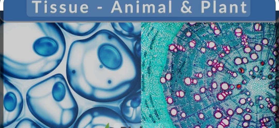
TISSUE ; A group of cell that are similar in structure and work together to achieve a particular function called Tissue ,
The microscopic study of tissue is called Histology .
PLANT TISSUE : MERISTEMATIC TISSUE / GROWTH TISSUE:
These are simple living tissues having thin walled compactly arranged immature cells which are capable of division and formation of new cells .
The main feature of meristematic tissues :
It has thin primary cell wall (cellulose )
In compact tissue the intercellular space are absent ,
generally vacuoles are absent , dense cytoplasm and prominent nuclei present
, Actively dividing cells are present in the growing regions of plants such as root and shoots tips
CLASSIFICATON OF TISSUE ON BASIS OF ORGIN
A) Primary Meristem/Pro meristem:
It is derived directly from the meristem of the embryo.
They consist of the cells derived from the primary meristem.
They add to primary growth of the plants.
B) Secondary Meristem ;
These are having cells derived from primary permanent tissue .
They usually add to the diameter of plants .
CLASSIFICATION of TISSUE ON THE BASIS OF LOCATION :
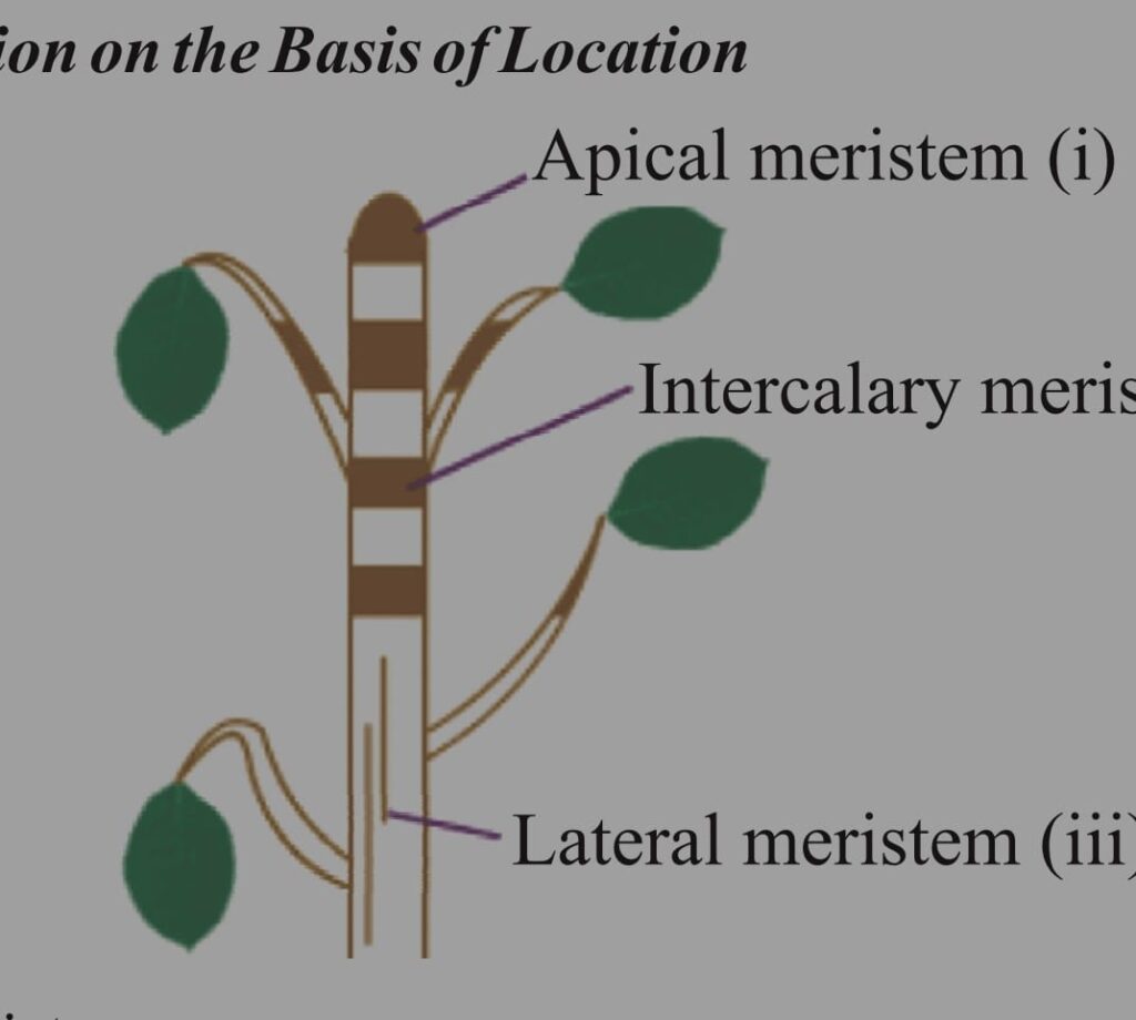
A) APICAL MERISTEM
It is present at the growth tips of stem and roots. Cell division in this tissue leads to the elongation of stem and roots thus it is involved in primary growth of the plant
B) INTERCALARY MERISTEM
It is present behind the apex , helps in longitudinal growth
.It is the part of meristem which is left behind during the growth of the period
These are present at the base of the leaf and internode region.
These lead to increase in the length of leaf
eg grass stem, bamboo stem , mint stem
C) LATERAL MERISTEM/CAMBIUM ;
It is also called as secondary meristem.
It occurs along the side of longitudinal axis of the plant,
It gives rise to the vascular tissues and responsible for the growth in girth of stem and root .
They are responsible for the secondary growth by increasing the grith.
PERMANENT TISSUE ;
The permanent tissue are formed from those meristematic cells which are left behind and have lost their capability to divide ,
The division and differentiation of the cells of meristematic tissue give rise to permanent tissues.
They have definite shape ,size , thickness they may be dead or living
As a result of cell differentiation the meristematic tissues tend to form different type of permanent tissues .
In cell differentiation , developing tissues changes from simple to more complex forms to perform various specialized functions.
Depending upon structure and composition the permanent tissues are classified into two types
A) SIMPLE PERMANENT TISSUE ( It is Supporting and protective tissue )
PROTECTIVE TISSUE;
These Dermal tissues are primarily protective in function .
They consist of
a) EPIDERMIS It forms one cell thick outermost layer of various body organs of plants such as leaves, flowers, stem and roots.
Epidermis is covered outside by cuticle .Cuticle is a water proof layer of waxy substance called cutin which is secreted by the epidermal cells and provide protection against the loss of water and also invasion by microbes.
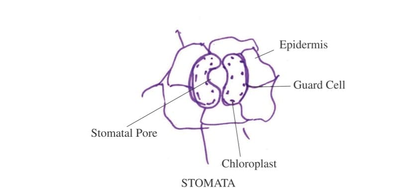
Cells of epidermis of leaves are not continuous at some places due to presence of small pores called stomata.
Each stomata is guarded by a pair of bean shaped cells called guard cells these are only epidermal cells which possess chloroplasts the rest being colourless.
FUNCTION of Epidermis :
The main function is to protects the plant from desiccation and infection .
The cuticle of the epidermis cuts the rate of transpiration and evaporation of water and prevent wilting of leaves .
stomata Function: It allows the gaseous exchange to occur during photosynthesis and respiration also helps in transpiration
PHELLEM/ CORK :
In older roots and stems , tissues at the periphery become cork cells.
They are made of dead cells with thick walls without any intercellular space
The cell wall in the cork deposit waxy substance suberin and it is impermeable to water and gases due to deposition of suberin
The cork cells are without any protoplasm but are filled with resin or tannins .

FUNCTIONS of Cork :
It is protective in function the cork cells prevent p lants from desiccation , infection and mechanical injury
It is perviousness , lightness , toughness , compressibility and elasticity so make the cork valuable for commercial use
SUPPORTING TISSUES
These are supportive in function and are of three types ;
Basic type of plant tissues are
Parenchyma Tissue,,
collenchyma Tissue ,
sclerenchyma tissue
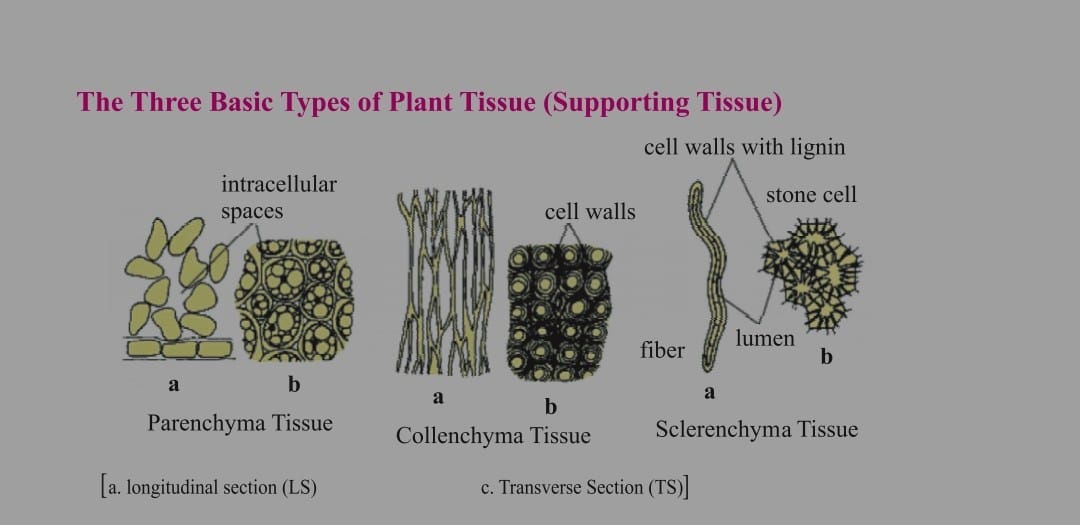
Parenchyma Tissue:
It is the fundamental backing tissues
They are loosely packed thin walled cells, oval / spherical in shape with the large space between them ,
Cell wall are composed of cellulose and pectin
large central vacuole for food and water storage are present
primary function is food storage and packing.
Parenchyma and it’s Type
a)Idioblast
some parenchyma involved in storage of excretory substances such as resin tannin gum and oil in typical Idioblast
In Typical Parenchyma Chlorophyll is absent
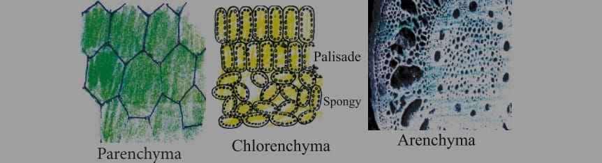
b) Chlorenchyma
They are the tissue containing chloroplast to perform photosynthesis ( eg: mesophyll cells of leaves )
c)Aerenchyma:
They are found in hydrophytic plants due to having space between them for which they have buoyancy for floating
COLLENCHYMA TISSUE :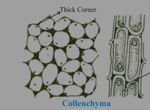
It is the living mechanical tissue.
It is elongated cell with thick corners,
localized cellulose and pectin thickening .
It provides flexibility and easy bending of various parts of plants .
few chloroplast may be present .
It give mechanical strength and elasticity to growing stems
It has very little intercellular space .
There are fibres in L.S
They are composed of extremely thick walled cells with little or no protoplasm
they are dead and possess very thick lignified walls
Lignin is water proof material
intercellular spaces absent.
SELERENCHYMA TISSUE :
They are of two types
a) Sclereids
These are also called grit cells / stone cells .
These are small cells where lumen is so small due to higher thickening of cell wall , as present in drupe fruit such as mango / coconut/ walnuts/legume seeds .
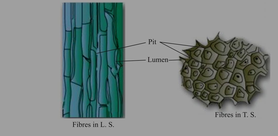
b) FIBRES
They are long ,narrow , thick lignified cells . Lumen is a large as compared to selereids . Generally 1-3 mm long .
In the thick walls of both the fiber and selereids thin aeras called as pits are present
Uses :
These are used in the manufacture of ropes, mats and certain textile fibres.
jute and coir are obtained from thick bundle of fibres .
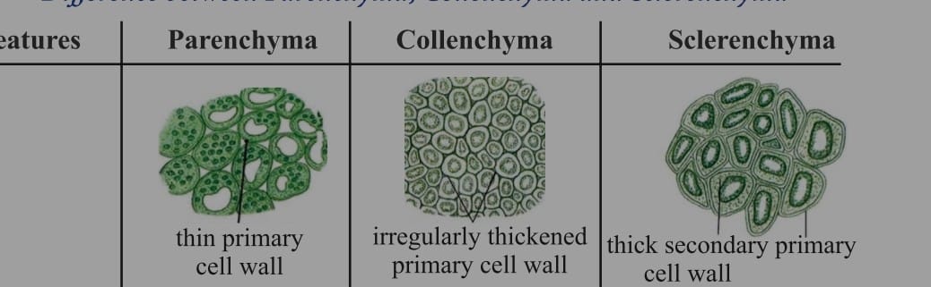
DIFFERENCE BETWEEN PARENCHYMA ,COLLENCHYMA AND SCLERENCHYMA TISSUE
A) Parenchyma Tissue ;
It has primary cell walls
The cell shape is isodiametric cell which are oval , spherical and polygonal in shape ,
Thin cellulosic cell wall , cytoplasm is abundant ,
Nucleus is present with large vacuole ,intercellular space is present
they are basically packing tissue ,all soft part plant pith , cortex and medullary rays
the main function is food storage, photosynthesis , provide buoyancy to hydrophytes
B) COLLENCHYMA TISSUE:
: Primary cell wall are irregularly thickness,
They are circular, oval or polyhedral in shape , cytoplasm is present , vacuolated , intercellular space absent ,
They are found in dicots stems, petiole and beneath the epidermis and
absent in monocot and roots
it provides tensile strength , mechanical support , photosynthesis
C) Sclerenchyma Tissue:
It is variable in shape . fibres and sclereids ,
having lignified secondary cell wall present ,
cytoplasm is absent having dead tissue, no vacuoles , no intercellular space. present in Dicot hypodermis, bundle sheath ,pericycle , seed, pulp of fruits
this protect the stress and strain mechanical strength.
COMPLEX PERMANENT TISSUES ;
It consist of more than one type of cells which work together as a unit
It helps in the transportation of organic materials, water and minerals
It is also known as conducting or Vascular tissue
Xylem and Phloem together form vascular bundles .
XYLEM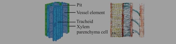
It is also known as wood and is a vascular and mechanical tissue.
It help in transportation of water and minerals from the soil to the plants parts.
Xylem is consists of
a) Tracheid: which are elongated dead cells /primitive elements mainly involved in the conduction of water and minerals in gymnosperms
b) Vessels:
They are advance element ( generally found in angiosperm )
Vessels are cylindrical tubelike structure place done above the other end to end which form the continuous channel for efficient conduction of water
c)Xylem Parenchyma :
They are small and thick walled parenchymatous cells designed for the storage of starch /food.
d) Xylem Sclerenchyma
They are non living fibers with thick walls and narrow cavities which provides mechanical support
Except xylem Parenchyma all other xylem elements are dead
The annual rings present in the trunk of a tree are xylem rings.
By contented number of annual rings we can determine the age of the plants
PHLOEM :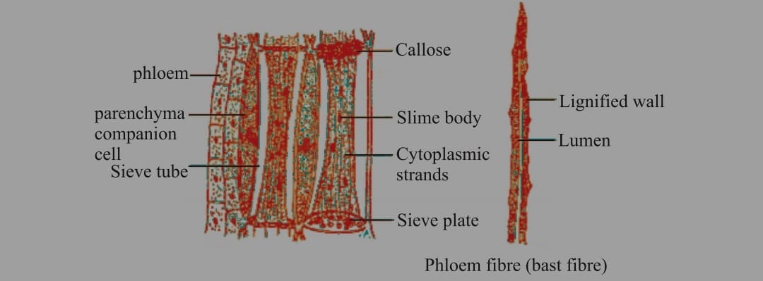
It transport /translocation food from leaves to other parts of the plant. All the phloem cells are living except phloem fibre.
It consist of four parts
a) Sieve tubes
They are the tabular structures’ made up of elongated, thin walled cells placed end to end .
The end walls of sieve tube cells are perforated by numerous the pores called sieve plates ,
The Nucleus of sieve cell degenerates at the maturity. However cytoplasm persists because of protoplasm continuation of sieve tube with the companion cell through plasmodesmata
b) Companion cells
They have dense cytoplasm and prominent nuclei
the sieve tubes and companion cells are also called sister cells because they originate from single mother cells
c) Phloem fibre/ Phloem Sclerenchyma
They give mechanical support to sieve tubes and dead
d)Phloem parenchyma
store food and help in radial conduction of food ,
Difference between Xylem and Phloem :
Xylem are dead cells, thick , having lignin , impermeable , carries water and minerals upward flow direction .
Phloem are living cells ,they are thin , permeable , having sieve plates it carries sugar and flow is upward and down ward
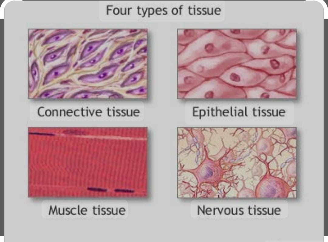
EPITHELIAL TISSUE ;
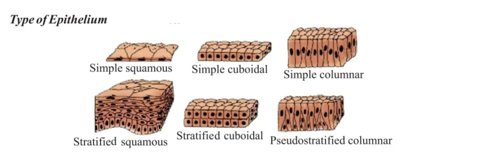
The cells of epithelium are set very close to each other tightly packed and the tissue rests on a non cellular basement membrane and consists of single layers of cells.
It covers all the organs and the line cavities of hollow organs like stomach .
It is primarily protective in function.
There are different types
A) SQUAMOUS EPITHELIUM :
It is also called pavement epithelium ,
, cells are arranged end to end likes tiles on the floor,
cells are polygonal in surface view
It forms the delicate lining of cavities ( mouth, oesophagus, nose , pericardium, alveoli )blood vessels and covering of the tongue and skin.
Epithelial cells are arranged in many layers to prevent wear and tear in skin . This pattern is stratified squamous epithelium.
B) CUBOIDAL EPITHELIUM :
They are cube like cells that fit closely , cells look like squares in the section but free surface appears hexagonal.
It is found in kidney tubules, thyroid vesicles and in the glands ( Salivary glands Sweat glands )
It form germinal epithelium of gonads (testes and ovaries )
It is involved in absorption, excretion and secretion It also provides mechanical support
C) CILIATED EPITHELIUM :
These cells may be cuboidal / columnar.
found in the respiratory tract , lining of the super duct, oviduct and kidney tubules
On its free surface are present protoplasmic outgrowth called cilia.
It helps in the movement of ova in fallopian tubes
D) GLANDULAR EPITHELIUM :
Gland cells secretes substances at the epithelial surface.
Sometimes position of epithelial tissue folds inward and form multicellular gland so it is called GLANDULAR EPITHELIAL
CONNECTIVE TISSUES
The cells of these tissue are widely spaced and embedded in an intercellular matrix.
Their basic function is to provide support to different organs and keeping them in place
Connective tissues have two components as matrix and cellular part.
A) FLUID or VASCULAR TISSUE ;
Blood and Lymph : Blood is a connective tissue,
fluid matrix of blood is plasma having wandering/ floating cells called corpuscles,
blood helps in the transportation of various materials such as nutritive substances , gases, excretory products and hormones etc.
i) Plasma
it form 55% part of blood, constitution 90-92% Water 7% Protein 0.9% ( Albumin , fibrinogen , globulin ) and inorganic salts
ii) Corpuscles
It forms 45% part of blood
iii) RBC (Red Blood Corpuscles )
they are also called erythrocytes, containing red color respiratory pigment called hemoglobin that helps in transportation of oxygen
iii) WBC ( White Blood Corpuscles ) /Leucocytes
they ae also called as soldiers of our body, it provide immunity .
They are irregular amoeboid ,phagocytes cells that protect our body by engulfing bacteria and other foreign particles they are of five types monocytes, Lymphocytes , Basophils , Neutrophils Eosinophils
iv) Blood platelets / thrombocytes :
They are spindle shaped cells which are involved in clotting of blood .
SKELETAL TISSUE :
a) BONE :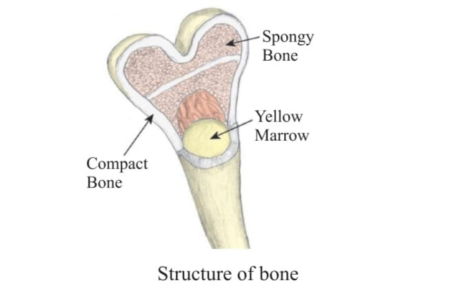
It is hard connective tissue that form supportive framework ie skeleton of our body .
It is of two type
i) Bone
the matrix of bone is very hard because of salts such as calcium , phosphate , CaCO3 (60-70%) and protein ossein .
Bone cells / osteoblast are embedded in the hard matrix .
Matrix is deposited in the form of connective layers of lamellae formed around a central canal , the bone cells occupy small spaces the concentric layers of matrix
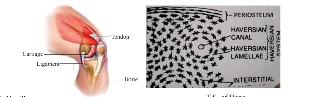
b) CARTILAGE
These tissue are elastic, less harder as compared to bones,
Elasticity is due to the presence of chondrin/protein. Cells are called as chondrocytes which are widely spaced and matrix is reinforced by fibres
Itis found at joint of bones of bones , in those , ear , trachea and larynx it provides the flexibility and great tensile strength.
FIBROUS TISSUE /Dense Regular Connective Tissue:
It is of two type
i) Ligament
ii)Tendon
It is the most abundant type of connective tissue
It is further divided into following types yellow fibrous connective tissue/ ligament
They are very elastic due to the presence of a network of yellow fibres in the matrix called ligaments. These ligament are attached to the bone to bone .
white fibrous connective tissue / tendon which have very little matrix containing abundant white fires forming layers and non elastic in nature
Bundles of these tissue are called tendons .These tendons attach muscles to bones .
Areolar Tissue :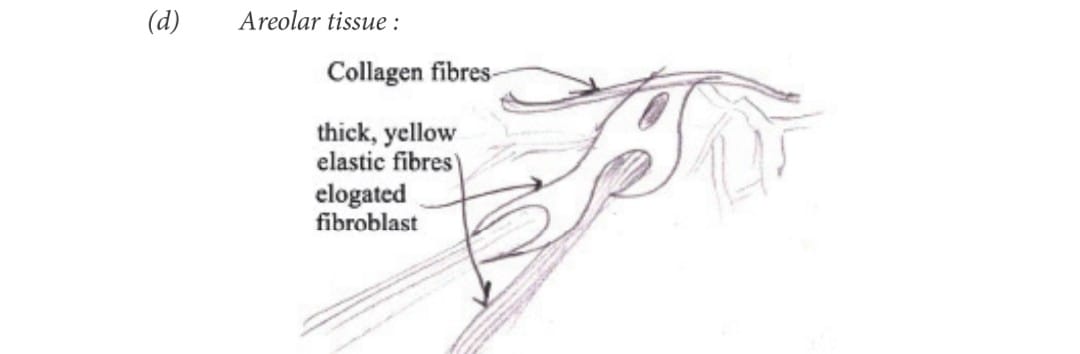
The tissue fill the space inside the organ and found between the skin and muscles around the blood vessels , nerves and in bone marrow .
It is a supporting and packing tissue .
It also helps in repair the tissue after injury
ADIPOSE TISSUE :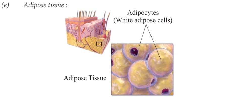
These are oval and round cells , filled with fat globules called adipocytes.
It is found in subcutaneous layer below the skin, around the heart, brain, below eye balls . It acts as insulator and prevent loss of heat from the body .
It severs as a fat reservoir and keeps the visceral organs in position.
MUSCULE TISSUE :
Movements are brought about in our body with the help of muscular tissue .
They are long fibre like cells called muscle fibes .
They are capable of contraction/ relaxation because they are up of contractile proteins.
Types of Muscular Tissue:
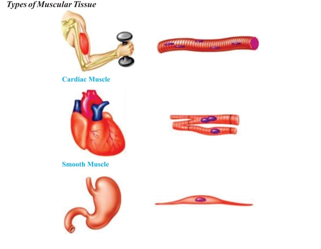
a) Skeletal Muscles :
These muscles shows alternate light and dark bands hence the name is striped or striated muscles.
They are also called as voluntary muscles because these are under the control of one’s will.
Muscle fibers/ cells are multinucleated and unbranched
Each fibre is enclosed by thin membrane which is called as sarcolemma
It’s Cytoplasm is called as sarcoplasm.
These muscles get tired and need rest.
b) Cardiac Muscle :
They are involuntary muscles only found in the walls of heart
They are unnucleated and branched. The branches are united by intercalated disc.
In these muscles rhythmic contraction and relaxation occurs throughout the life and never get tired .
c) Non Striated Muscles :
They are involuntary muscles also called as smooth muscles,
They donot show any alternate light and dark bands.
These muscles fibres are unnucleated and spindle shaped.
They are not enclosed by membrane but many fibres are joined together in the bundles. They constitute internal organs.
Such muscles are found in the walls of stomach, intestine, urinary bladder , bronchi ,iris of eye.
Peristaltic movement in the alimentary canal . are brought about by smooth muscles.
Difference between Striated, Non- Striated and Cradic Muscles
STRIATED MUSCLES :
They are present in limbs, body walls , tongue pharynx and the beginning of oesophagus.
They are cylindrical with fibres unbranched, multinucleated.
They are bounded by sarocolemma having light and dark bands ,
There is no oblique bridges and intercoalated discs ,
Nerve supply from central Nervos system
blood supply to it is abundant ,
have rapid contraction
They soon get fatigue used in voluntary action .
NON STRIATED MUSCLES :
they are found in posterior part of oesophagus , urino- genital tract, urinary bladder , iris of eye , dermis of skin and arrector pili muscles of hair etc
They are spindle in shape with unbranched fibres,
uninucleated ,
bounded with plasmalemma,
they do not have light or dark band
no oblique bridges and intercalated discs.
Nerve supply to it is from autonomic nervous system
The blood supply is scanty
having slow contraction ,
involuntary so do not get fatigue.
CARDIC MUSCLES :
They are present in the walls of heart , pulmonary veins and superior vena cava .
They are cylindrical in shape , having fibrous branch , uni- nucleate
bounded by sarcolemma ,
having faint light and dark bands
oblique bridges and intercalated discs present
Nerve supply from the brain and autonomic nervous system
, blood supply to it is abundant
having rapid contraction ,
never get fatigued and involuntary in nature .
NERVOUS TISSUE :
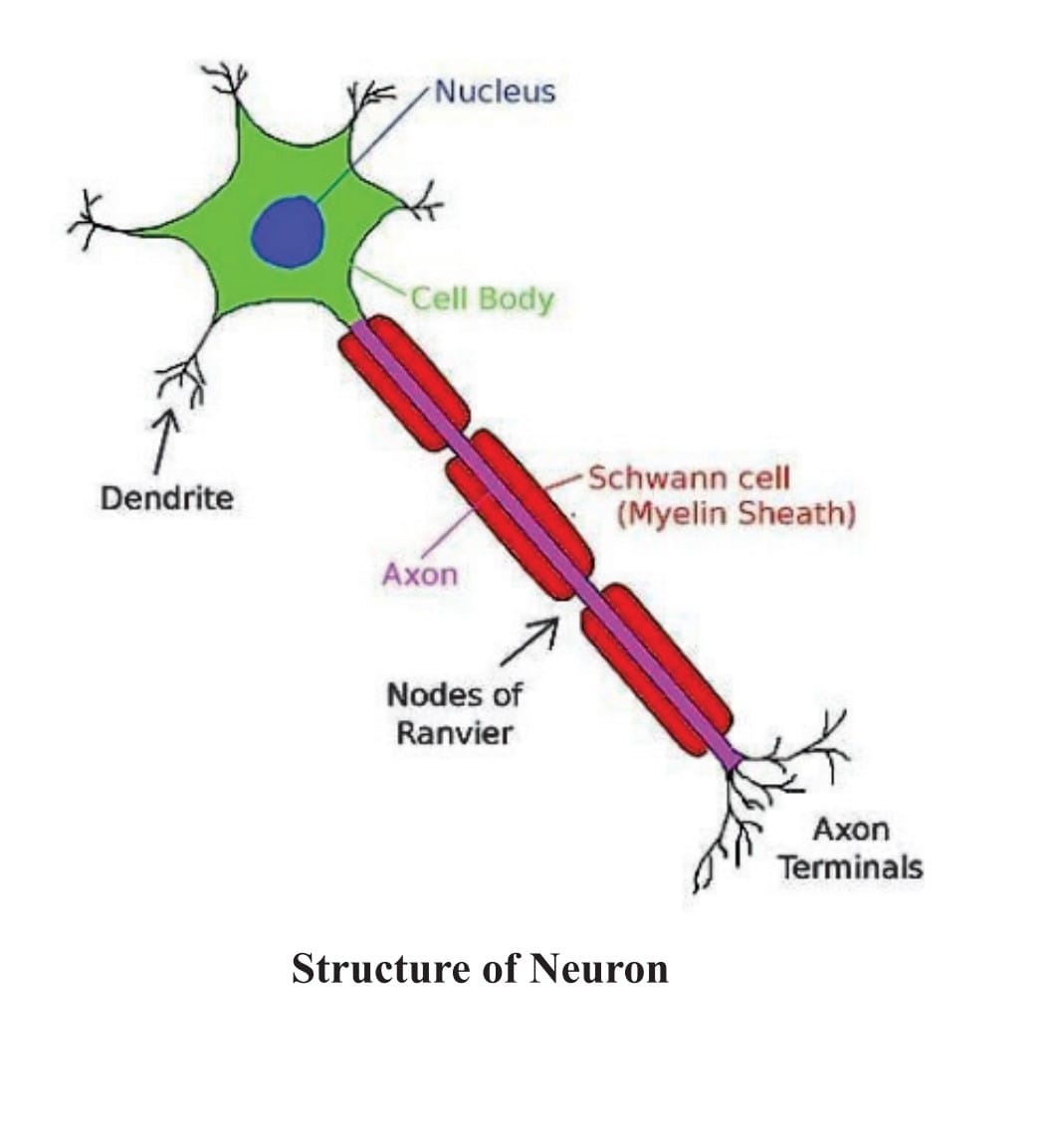
They are highly specialized tissues due to which the animals are able to perceive and respond to the stimuli .
Their functional unit is called nerve cell/ neuron.
The main part of Neuron
cell body is cyton covered with plasma membrane.,
short hair like extension rising from cyton are dendrons which are further which are further subdivide into dendrites .
Axon is long, tail like cylindrical structure with fine branch at the end and is covered by sheath , which is known as myelin sheath.
Nerve ending of one neuron is very closely placed to dendron’s of the other neuron to carry impulses from one neuron to another in the form of electro- chemical waves. This closes the proximity is called synapse
Read More : Fundamental unit of life cell
Follows us on : Facebook